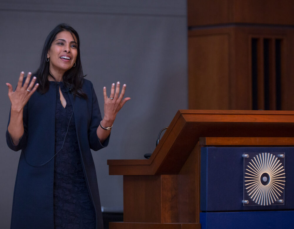
Sangeeta Bhatia works at the intersection of medicine and engineering, using nanotechnology—the “tiny technologies”—to develop tools that can diagnose cancer, liver diseases, and other conditions without biopsies or other invasive procedures. As a graduate student Bhatia pioneered a method of growing mini-livers that are used by pharmaceutical companies to test medicines. At the MIT lab she now heads, they are investigating how to grow liver tissue to help people with hemophilia and other diseases. She has also studied ways to use nanotechnology to deliver chemotherapy, immunotherapy, and gene therapies to tumors; to monitor how well medicines are working in patients; and to kill antibiotic-resistant bacteria.
In May 2019 she visited the Science History Institute to accept the Othmer Gold Medal. Distillations writer Meir Rinde sat down with Bhatia to find out how nanodiagnostics work, what impact they could have in developing countries, and why she adores the liver. What follows are excerpts from their discussion, condensed and edited for clarity.
Using nanoparticles to screen for disease
These teeny, tiny particles [about a thousand times narrower than a human hair] can be injected in the bloodstream and circulate in your body looking for disease. We make them responsive to certain enzymes that are disease associated. When the particle finds the enzyme that it’s been designed for, there’s a chemical reaction and they emit a little reporter, a little synthetic signal, which is a chemical molecule that’s not found in your body. That molecule finds its way into the urine after about an hour. The patient gets a shot, the molecules roam and look for disease, and then an hour later you do a urine test. If you find these signals in the urine, then you could diagnose this disease.
There are about 550 enzymes in the family [of enzymes] that we’re measuring. They are connected to almost every kind of disease: cancer, infection, inflammation, blood clotting.
In the lab we realized we could make panels [that test for 10 to 20 conditions at once]. So far we’ve done about a dozen diseases, and it seems to work pretty well. About three years ago we realized that we should take this to patients. We did that by starting a company called Glympse. They are going to start clinical trials [in the third quarter of 2019]. We’re really excited to see how this plays out in the marketplace.
We actually invented [nanosensors] by accident. We weren’t trying to invent sensors that would come out in the urine. We were trying to make nanoparticles that would be smart contrast agents: when you go to get an MRI scan, it would give you some functional information. It so happened when we were doing the experiments with mice that had tumors, the urine was lighting up. We had this “aha” moment, like, “Oh, we don’t need an imager at all. We can do a noninvasive test.”
On her long-standing interest in the liver
The title of my talk tonight is “An Engineer’s Love Affair with the Liver.” I like to say that I fell in love with the liver the first day of graduate school, and it’s been a lifelong journey. I just was never done with it. It’s vital for life, it has 500 functions, it can regenerate without a stem cell. As an innovator, what’s drawn me to it and kept me with it is that it’s a field of medicine where we have shockingly little to offer. That’s in comparison, let’s say, to cardiology, where we have bypass, we have stents, we have medicines, we have a whole host of interventions. In the liver really all we have is transplant, beginning and end of story. That makes it feel like anything you do would be potentially impactful.
It’s been an area that has long been under-studied because it’s hard [to research], and it’s deep in the body, but also for all kinds of social reasons here in the West. [Liver ailments were] seen for a long time as a class of diseases that were self-inflicted. So [there’s] alcohol, and then there’s hepatitis, which is blood-borne. For the first time the liver’s really having its heyday. Gene therapy—CRISPR, genome editing, RNA interference—can very readily be delivered to the liver. Now everyone’s really interested in liver disease. I’ve been working in this field for 20 years and nobody cared. All of a sudden everybody’s at my party, which is great.
I have two daughters, and my oldest daughter is taking biology. She’s in her sophomore year in high school. I was looking at her book to see what does it say about the liver, which is the first thing I always ask. And it’s like one paragraph on one page, and it says vague things like “metabolism.” Even the way we teach it is so uninteresting. I can see why it hasn’t drawn attention over the years.
The role nanoparticle screening could play in patient care around the world
One of the diseases that we’re really interested in is NASH, nonalcoholic steatohepatitis. There’s fat accumulation in the liver, the liver gets damaged, and eventually it scars. Right now the only way to test for that is to do a biopsy. That’s a full-day procedure that’s got some finite complication rate, and it turns out that all of these enzymes that we’re capable of measuring are involved with that disease.
We also have a test for blood clotting. We have a test that we’re developing for pneumonia to see if you have bacterial or viral pneumonia. We have a test to see if in the setting of prostate cancer, is it aggressive prostate cancer or slow-growing prostate cancer?
One of the most appealing use cases that we’ve imagined is that you could have an oral formulation, a pill—which we don’t have yet, but we’re working on—and then a urine test that would have a paper-strip readout. At the point of care and in the absence of clinical infrastructure, you could do really high-end molecular diagnostics. That starts to get you into thinking about how transformative diagnostics could be around the world, where we don’t have the infrastructure that we have here. Cancer screening is an example. If there is no colonoscopy, no mammogram, and no Pap smear available, can you imagine doing a urine test on a paper strip and taking a picture with a smartphone and sending it to another provider? For us it’s been really exciting to think about how to point this to the biggest unmet medical needs.
I’m of Indian origin. When I was growing up, we used to spend summers in India. An aunt [who] was a physician would take me to the clinic, and I would see what medical care is like in a low-resource setting. That stuck with me in the back of my head. I’ve always wanted to get back to inventions that could make an impact.
How nanosensors can help avoid overdiagnosis and unnecessary intervention
One of the use cases we’ve been really interested in is, in the setting of a potential cancer, can you stratify whether it’s cancer or not? Furthermore, could you tell, if it is cancer, is it an aggressive cancer that needs treatment or is it a slow-growing tumor that you would die with but not die from? We’ve been working on two different versions of those use cases. The first one is in lung cancer. The new recommendations that have come out from the U.S. Preventive Services Task Force are that patients at risk for lung cancer because of smoking histories should be screened by low-dose CT scanning. That’s starting to happen, and it turns out that there are lots of things in the lungs that look like they could be cancer, but they’re not, especially in the lungs of long-term smokers. What happens then is you get a bronchoscopy and a sampling of that lesion, and 95% of the time it’s a false positive.
It’s not even an early detection question. It’s a question of, in the setting of a scan that’s positive, can you just do a urine test and see if there is a cancer or not? That would lead to less intervention, right?
Similarly, in prostate cancer, what happens today is, you typically get biopsied, and [there are high, low, and] intermediate scores. They’re called Gleason scores. So with a Gleason [intermediate score of] six or seven, you don’t know [what to do]. You together with your clinician make a judgment call based on your age and risk tolerance and all kinds of things about whether you would do surgery or intervene.
We published a paper last year showing that the enzyme profile of an aggressive cancer is different than the slow-growing one. The hypothesis was that, basically, as a tumor grows and invades the tissue around it, it has to make these enzymes to kind of chew its way out.
The challenges of bringing new diagnostic technologies to market
We talked here at the Institute this afternoon a lot about financial models. One challenge with diagnostics in general is that they’re actually really hard to translate to patients. They’re hard to commercialize, they’re hard to get funded, they’re hard to get reimbursed for. And the screening applications are the hardest of all because you have to do a great big study in patients who are at risk to see whether your test works or not. We’d like to get there someday, but if you really want to see your tool make an impact, you also have to identify near-term wins, threading the needle between being the visionary and the pragmatist.
Funding new technologies versus increasing preventive care in global oncology
We do a lot of work with the AIDS Foundation, which does not have global oncology as a strategic priority. That’s not in their mandate. Last year they asked me to give a spotlight talk about some of the work we do, and they asked me to be provocative. I don’t know that they expected me to get up there and give basically a TED-like talk about how they should be investing in global oncology.
Global oncology shares many attributes with the HIV story. If you look at what everyone said about HIV 20 years ago, it’s exactly the same. “Oh, we can’t treat it, we don’t have the right medicines, we have to fix the medical infrastructure problem in order to be able to address it. There are many lifestyle issues that feed into this. It’s too complicated.” And now look where we are. The cost of medicines has come down by 99%. In my view, looking forward, we absolutely have to take the same approach [with global oncology].
But you have to develop [the technologies] for a low-resource setting. If you think about what the cancer establishment here in the U.S.—and I’m part of it—is doing . . . we’re all very excited about immunotherapy, and we are screening with expensive instruments, and now we’re really excited about liquid biopsy, which is DNA sequencing. But if 70% of the cancer burden by 2025 is going to be in low- and middle-income countries, none of those [technologies] are going to apply in that setting very soon. What about a grand challenge where you actually just need a CT scanner that can operate on intermittent power, that never needs to be serviced? There are some very fundamental inventions that probably could transform the landscape that nobody’s working on. I’ve been really interested in how our tools could feed into that environment. What people have said to me is, how can you make a cancer diagnostic for a country or a setting where there’s no surgical suite? All I can say as the inventor is, you can’t not invent it! You have to invent for the future that you hope is going to happen.
Growing liver tissue and its clinical potentials
What we’ve been really interested in is [whether we can] position liver cells using some 3-D printing techniques so that if you implant them, they would recruit blood vessels and they would grow. The reason we knew they should be able to grow is because the liver can regenerate, unlike other tissues. Those are the experiments we’ve been doing, and it’s working. In mice we can get them to spontaneously vascularize. We put [the liver cells] in just the right arrangement and they grow about 50 times in the setting of a liver injury. We are just now starting to think about [taking] this to patients to do a start-up.
There’s a class of liver diseases where you don’t need a whole transplant to fix them. Most of them are metabolic liver diseases. Those are diseases that you could fix with 1% to 2% of the liver mass. Hemophilia is an example. You get to therapeutic levels of some of the clotting proteins without needing a whole new liver. For some of these applications our idea’s not to replace the liver but to make a little satellite liver. So that’s what we would start with.
The many aspects of leadership
To some extent you’re like the ultimate entrepreneur when you’re an academic. You have to have the vision, you have to raise the money, you hire the talent, you create the culture, you help prioritize ideas and interactions. And then I consider it to be a part of my role to be a mentor. My goal always is to create independent thinkers. I tell all my students, by the time that you graduate, the perfect trajectory is that now you know more than I do, and you go off and you create your own world. I don’t have a lab of people who are doing my bidding. I’m really trying to create independent scholars.
The great thing about being an academic is that you get to evolve your role. I have chosen issues to advocate for what I care about. I care a lot about diversity—so women in STEM, at every level—and I work and speak about that. I’ve been in the last few years very interested in translation because we have enough inventions that I feel like, “How can I get these into patients?”


